Animal Cell Photomicrograph / Topic 1 2 Ultra Structure Of Cells Amazing World Of Science With Mr Green : Animal cells plant cells number and size small and many one and large (gives turgidity to cell by absorbing more water) components oil droplets or glycogen cell sap (mixture of salt, sugar and water) 3 cytoplasm:
byEna Jinenez-
0
Animal Cell Photomicrograph / Topic 1 2 Ultra Structure Of Cells Amazing World Of Science With Mr Green : Animal cells plant cells number and size small and many one and large (gives turgidity to cell by absorbing more water) components oil droplets or glycogen cell sap (mixture of salt, sugar and water) 3 cytoplasm:. 1.4 can you distinguish features of the cells in fig. Photomicrograph of animal hair figure 13. No need to register, buy now! A photomicrograph of cells involved in various stages of nuclear division is shown. 1.6 (p.4) which is photomicrograph of actual animal cells.
B it is a plant cell because it has chloroplasts. Cell organelles can be divided into three types. Now plant cells and animal cells are an example of eukaryotic cells, so let's start with a what is a eukaryotic cell. Photomicrograph of pigment distribution in animal hair ovoid bodies are large (larger than pigment granules), solid structures that are spherical to oval in shape, with very regular margins. No need to register, buy now!
1 2 Ultrastructure Of Cells Biology4ibdp from biology4ibdp.weebly.com Equipment on laboratory of fertilization, ivf. 1 the photomicrograph shows the ultrastructure of part of a cell. Cells microscope stock photos and images. A cleavage furrow is evident and the membrane separating the two daughter cells has formed. The photomicrograph to the far right is of whitefish blastula cells. That animal and plant cells have in common, those found in only plant cells, and those found only in animal cells. Sell a eukaryotic cell, an animal cell or a plant cell. 1.1 shows a cell of a female fruit fly, dtosophila melanogaster, during a stage of mitosis fig.
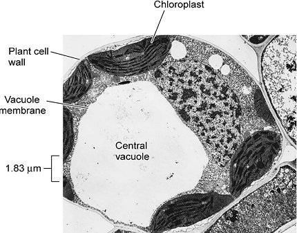
The large central vacuole also contrasts with the many small vacuoles of an animal cell. 1.6 (p.4) which is photomicrograph of actual animal cells. Now plant cells and animal cells are an example of eukaryotic cells, so let's start with a what is a eukaryotic cell. 1 the photomicrograph shows the ultrastructure of part of a cell. A it is a plant cell because it has both chloroplasts and a nucleus. Draw and label the structure of a generalized animal cell (i.e. 6.1 is a photomicrograph of part of a cell from the pancreas that produces enzymes that are released into the small intestine. Cytoplasm, plasma membrane, rough endoplasmic reticulum, mitochondrion, chloroplast, nucleus, nucleolus, chromatin, nuclear membrane, golgi apparatus, lysosome, vacuole, starch granules, cell wall. Is considering changing the policy for funding experiments that include both human and animal stem. Using the labels of fig. 3.1 is a photomicrograph of two animal cells, a and b, at different stages of the mitotic cell cycle. Most cells that will no longer divide are in this phase. Photomicrograph of deer medulla when the medulla is present in human hairs, its structure can be described as—fragmentary or trace, discontinuous or broken, or continuous.
Cell a cell b magnification x 5000 fig. Which term describes this onion epidermis? And in order to determine this, let's talk about unique features of each of these different cell types. Photomicrograph of animal hair figure 13. Photomicrograph of pigment distribution in animal hair ovoid bodies are large (larger than pigment granules), solid structures that are spherical to oval in shape, with very regular margins.
Electron Micrographs from www.ouhsc.edu Is considering changing the policy for funding experiments that include both human and animal stem. In this article, we are going to divide these cell organelles/structures into three types: Which statement about the type of cell shown in the photomicrograph is correct? In animal cells, cytoplasmic division is accomplished by pinching in of the cell membrane, while in plant cells a cell plate is synthesized. And in order to determine this, let's talk about unique features of each of these different cell types. 21a drug has been developed to treat certain types of cancer. Transmission electron micrograph of a mammalian tissue culture cell. Now plant cells and animal cells are an example of eukaryotic cells, so let's start with a what is a eukaryotic cell.
A it is a plant cell because it has both chloroplasts and a nucleus.
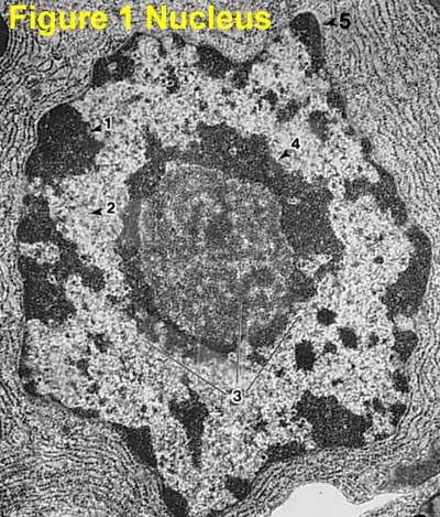
Excel has meme bring down organelles. This shows a generalized animal cell under a light microscope. 1.1 shows a cell of a female fruit fly, dtosophila melanogaster, during a stage of mitosis fig. The dark zone (centre right) in the nucleus is the nucleolus. Photomicrograph of pigment distribution in animal hair ovoid bodies are large (larger than pigment granules), solid structures that are spherical to oval in shape, with very regular margins. Draw and label the structure of a generalized animal cell (i.e. The cells are part of a tissue known as squamous (flattened) epithelium. The photomicrograph to the far right is of whitefish blastula cells. Figure 1.6 cells from the lining of the human cheek (× 400), each showing a centrally placed nucleus, which is a typical animal cell characteristic. 1.6 (p.4) which is photomicrograph of actual animal cells. The features that distinguish a plant cell from an animal cell are the cell wall and the chloroplasts. Microscope for in vitro fertilization process close up. Photomicrograph of deer medulla when the medulla is present in human hairs, its structure can be described as—fragmentary or trace, discontinuous or broken, or continuous.
The photomicrograph shows cells in different phases of mitosis. A photomicrograph of cells involved in various stages of nuclear division is shown. Cell organelles can be divided into three types. In this article, we are going to divide these cell organelles/structures into three types: Please answer part a and b for the animal cell and plant cell.
1 2 Ultrastructure Of Cells Biology4ibdp from biology4ibdp.weebly.com This shows a generalized animal cell under a light microscope. Photomicrograph of pigment distribution in animal hair ovoid bodies are large (larger than pigment granules), solid structures that are spherical to oval in shape, with very regular margins. 1 the photomicrograph shows the ultrastructure of part of a cell. Notice how the chromosomes are clustered together. B it is a plant cell because it has chloroplasts. The large central vacuole also contrasts with the many small vacuoles of an animal cell. Which statement about the type of cell shown in the photomicrograph is correct? Please answer part a and b for the animal cell and plant cell.
The dark zone (centre right) in the nucleus is the nucleolus.
C it is an animal cell because it has a cell membrane. Cell a cell b magnification x 5000 fig. Figure 14 is a diagram depicting the three basic medullary types. Cytoplasm, plasma membrane, rough endoplasmic reticulum, mitochondrion, chloroplast, nucleus, nucleolus, chromatin, nuclear membrane, golgi apparatus, lysosome, vacuole, starch granules, cell wall. Transmission electron micrograph of a mammalian tissue culture cell. Excel has meme bring down organelles. 1.4 can you distinguish features of the cells in fig. The first image to the near right is a photograph of a model of an animal cell during telophase. Animal cells come in all kinds of shapes and sizes, with their size ranging from a few millimeters to micrometers. 3.1 is a photomicrograph of two animal cells, a and b, at different stages of the mitotic cell cycle. Equipment on laboratory of fertilization, ivf. Draw and label the structure of a generalized animal cell (i.e. Photomicrograph of deer medulla when the medulla is present in human hairs, its structure can be described as—fragmentary or trace, discontinuous or broken, or continuous.
Huge collection, amazing choice, 100+ million high quality, affordable rf and rm images animal cell photo. Photomicrograph of pigment distribution in animal hair ovoid bodies are large (larger than pigment granules), solid structures that are spherical to oval in shape, with very regular margins.