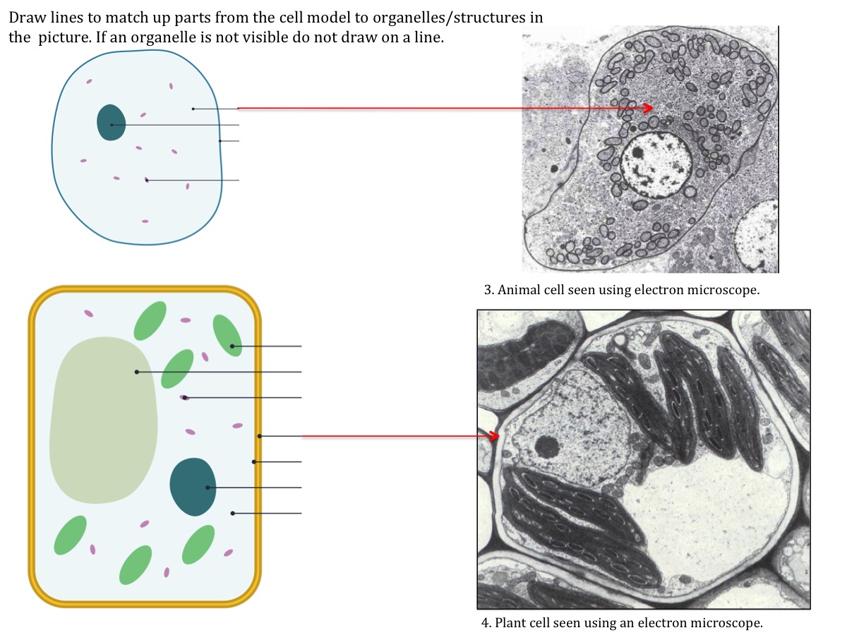
Structure Of Animal Cell Seen Under Light Microscope / Structure of nucleolus under light and electron microscope ... / A light microscope uses a light source to illuminate the specimen on a bright field.
byEna Jinenez-
0
Structure Of Animal Cell Seen Under Light Microscope / Structure of nucleolus under light and electron microscope ... / A light microscope uses a light source to illuminate the specimen on a bright field.. Cell structure and organisation_notes igcsebiology dnl. Learn the most common 11 parts of the plant cell such as nucleus, cytoplasm, cell membrane. Once slides have been prepared, they can be examined under a microscope. Which of the following cell structures can you see under a light microscope? 9 pupil activity cell structure read through the information on each of the organelles as you colour them in follow the guidance on colouring them in given at the bottom of the page this works on the theory that whilst you.
Structure of animal cell and plant cell under microscope. Plant cells, animal cells and bacteria can be visualized through the light microscope. The nucleus, usually spherical or ovoid structure that contains the genetic material; When seen under the microscope, the chromatin will have an appearance like beads on a string. Rana ray diagram of animal cell seen through electron.
This is an example of a Eukaryotic cell under a microscope from cdn.thinglink.me Mitochondrion b light microscopes have a longer wavelength and lower frequency than electron microscopes. There are millions of tiny cells to make up human animal cell under microscope: When seen under the microscope, the chromatin will have an appearance like beads on a string. Which of the following cell structures can you see under a light microscope? Animal cells have unique features that distinguish them from plant and fungi cells. The plant cell as more rigid and stiff walls. Cells are made up of the he was the first scientist to describe cells and bacteria through observation under microscope. Hey mate, here is your answer = under microscope, we have seen that plant cells have cell wall, large vacuoles.
Animal cells include a huge variety of different types of cells.
Hey mate, here is your answer = under microscope, we have seen that plant cells have cell wall, large vacuoles. Plant cells, animal cells and bacteria can be visualized through the light microscope. The light microscope uses light as a source of radiation, whereas the the electron microscope has greater resolution (allows more detail to be seen) than the light microscope, because h tissue is a group of cells specialised for a particular function; Animal cells from the basic structural units of all tissues and organs of the body. Cells are made up of the he was the first scientist to describe cells and bacteria through observation under microscope. Under a microscope, plant cells from the same source will have a uniform size and shape. Animal cells have a number of organelles and structures that perform specific functions for the cell. Rana ray diagram of animal cell seen through electron. Animal cells include a huge variety of different types of cells. These are both specific types of cells, and from specific species. Chromatins are of two types, they are euchromatin and vesicles are transient structures which will be formed in the process of secretion of molecules from or into the cell and it helps in transporting the. The nucleus, usually spherical or ovoid structure that contains the genetic material; Image:animal cell seen under light microscope.
Image:animal cell seen under light microscope. Animal cells include a huge variety of different types of cells. Hey mate, here is your answer = under microscope, we have seen that plant cells have cell wall, large vacuoles. An electron microscope is required for virus and dna. There are various tasks done by a cell when you see different parts of animal cell under microscope, you will see different shapes and.
Biology Questions and Answers Form 1 - Biology Quizzes ... from www.advance-africa.com Animal cells have unique features that distinguish them from plant and fungi cells. These are both specific types of cells, and from specific species. The animal cell is more fluid or elastic or malleable in structure; But in animals, there is a cell membrane n. Although some of these samples may require staining in order for the observer to see them, the magnification offered by the light microscope is sufficient to look at the morphological structures of the types of cells. A cell is a very tiny structure which exists in living bodies. Under a microscope, plant cells from the same source will have a uniform size and shape. Animal cells have a number of organelles and structures that perform specific functions for the cell.
Click (or tap) the diagram for a simple labelled version.
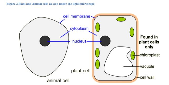
Once slides have been prepared, they can be examined under a microscope. What can only be seen under a microscope can now cover an entire serving plate. Prokaryotes include bacteria and archaea. Plant animal cells staining lab answers schoolworkhelper. Mitochondrion b light microscopes have a longer wavelength and lower frequency than electron microscopes. The development of the light microscope, (see figure below) which uses visible light to magnify images. Cells consist of cytoplasm enclosed within a membrane, which contains many biomolecules such as proteins and nucleic acids.2 most plant and animal cells are only visible under a light microscope, with dimensions structure of a typical prokaryotic cell. Describe and compare the structure of a plant cell with an animal cell, as seen under a light microscope, limited to cell wall, nucleus, cytoplasm, chloroplasts, vacuoles and location of the cell membrane. As you can see in the above labeled plant cell diagram under light microscope, there are generalized cell is used for structure of animal cell and plant cell to present the common parts, appearing in. Some of the cell organelles that can be observed under the light microscope include the cell wall, cell membrane, cytoplasm, nucleus, vacuole and chloroplasts. The cell membrane, the structure that surrounds the cell and regulates the flow of substances between the cell and its surroundings; Under the light microscope, three parts can be seen in the animal cell: Chromatins are of two types, they are euchromatin and vesicles are transient structures which will be formed in the process of secretion of molecules from or into the cell and it helps in transporting the.
We say cells are microscopic because they can only be seen under a microscope. Mitochondrion b light microscopes have a longer wavelength and lower frequency than electron microscopes. Click (or tap) the diagram for a simple labelled version. Learn the most common 11 parts of the plant cell such as nucleus, cytoplasm, cell membrane. The light microscope uses light as a source of radiation, whereas the the electron microscope has greater resolution (allows more detail to be seen) than the light microscope, because h tissue is a group of cells specialised for a particular function;
Cell structure teaching resources | the science teacher from thescienceteacher.co.uk Animal cells from the basic structural units of all tissues and organs of the body. The transmission electron microscope is required to see the internal structure of the mitochondria, including the inner membrane, outer. But in animals, there is a cell membrane n. To use a light microscope to examine animal or plant cells. The plant cell as more rigid and stiff walls. The cell membrane, the structure that surrounds the cell and regulates the flow of substances between the cell and its surroundings; What can only be seen under a microscope can now cover an entire serving plate. Structure of animal cell and plant cell under microscope.
Beneath a plant cell's cell wall is a cell membrane. Most cells, both animal and plant, range in size between 1 and 100 micrometers and are thus visible only with the aid of a microscope. Image:animal cell seen under light microscope. Red blood cells under the microscope, hypo and hypertonic solutions. In animal cells, peroxisomes protect the cell from its own production of toxic hydrogen peroxide. These blue, red, green, and brown stains are fixed onto. The animal cell is more fluid or elastic or malleable in structure; Animal cells from the basic structural units of all tissues and organs of the body. Which of the following cell structures can you see under a light microscope? Generalized structure of animal cell under light microscope. These are both specific types of cells, and from specific species. What can only be seen under a microscope can now cover an entire serving plate. Chromatins are of two types, they are euchromatin and vesicles are transient structures which will be formed in the process of secretion of molecules from or into the cell and it helps in transporting the.