Animal Cell Under Electron Microscope : Structure Of Animal Cell And Plant Cell Under Microscope Diagrams Animal Cell Cell Diagram Plant Cell Diagram : An electron microscope is a microscope that uses a beam of accelerated electrons as a source of illumination.
byEna Jinenez-
0
Animal Cell Under Electron Microscope : Structure Of Animal Cell And Plant Cell Under Microscope Diagrams Animal Cell Cell Diagram Plant Cell Diagram : An electron microscope is a microscope that uses a beam of accelerated electrons as a source of illumination.. Ishita observed a slide of eukaryotic cell under electron microscope. Can people see eukaryotic cells under a scanning electron. Most cells, both animal and plant, range in size between 1 and 100 micrometers and are thus visible only with the aid of a microscope. Cell membrane dr jastrow s electron microscopic atlas. Resolving power is the ability to distinguish between separate things which are close to each other.
When ultraviolet light hits an object, it excites the electrons of the object, and they give off light in. How is it different from animal cell? How to prepare a wet mount of cheek cells. 11 best electron microscope images in cell images on. What does an animal cell look like under an electron.
Animal Cell High Resolution Stock Photography And Images Alamy from c8.alamy.com Detail study of animal cell under electron microscope. Human skin under microscope 400x. How is it different from animal cell? However, the internal structure and organelles are more or blood cells are cellular structures found suspended in the plasma of the blood. Here's a diagram of a plant cell: Cell membrane dr jastrow s electron microscopic atlas. Under the microscope, animal cells appear different based on the type of the cell. Cytoplasm, ribosomes, rough endoplasmic reticulum;
1st john 1:1 holy hydrogen light of creation has been discovered glowing within the human cell wall plasma nucleus as seen with an electron microscope in.
Ishita observed a slide of eukaryotic cell under electron microscope. Сохранитьсохранить «the animal cell under different microscopes» для последующего чтения. What does an animal cell look like under an electron. Most cells, both animal and plant, range in size between 1 and 100 micrometers and are thus visible only with the aid of a microscope. However, they usually can achieve a maximum of 2000x magnification which is not sufficient to see many other tiny organelles. You see that many features are in common. Plant cell has cell wall and cell membrane and animal cell has vacuole and nucleus. When ultraviolet light hits an object, it excites the electrons of the object, and they give off light in. Detail study of animal cell under electron microscope. How is it different from animal cell? A cell is a very tiny structure which exists in living bodies. 4 main components of the cytoplasm (with diagram). The role and function of the plasma membrane;
11 best electron microscope images in cell images on. Here's a diagram of a plant cell: When you look at animal or plant cells under the electron microscope, you. Animal cell (as seen under electron microscope). Difference between animal and plant cell.
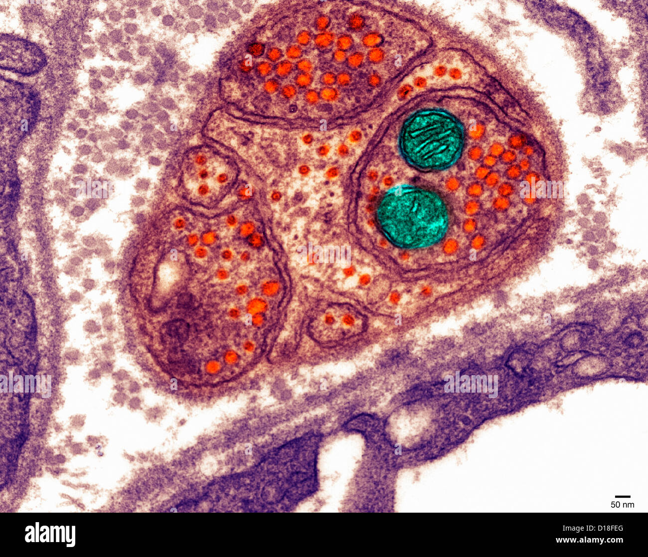
Animal Cell High Resolution Stock Photography And Images Alamy from c8.alamy.com There are two types of electron microscope 7 ultrastructure of an animal cell as seen through an electron microscope. Electron microscopes use electron beams focused by electromagnets to magnify and resolve microscopic specimens. Human skin under microscope 400x. You see that many features are in common. Electron microscope uses electrons and an ordinary microscope under a light microscope, the parts of a simple animal cell (e.g. Animal cell (as seen under electron microscope). Here's a photo of a plant cell under an electron microscope.
The animal cell is more.
Cautionary labels are given for products or containers containing hazardous material. Cytoplasm, ribosomes, rough endoplasmic reticulum; When you look at animal or plant cells under the electron microscope, you. What does an animal cell look like under an electron. Electron microscope uses electrons and an ordinary microscope under a light microscope, the parts of a simple animal cell (e.g. Human blood contains a number of blood cells on the basis of their purpose. Electron microscopy of cultured epidermal ebs 2117 cells reveals. That cells can be of different shapes and sizes. After completing this section, you should know: However, they usually can achieve a maximum of 2000x magnification which is not sufficient to see many other tiny organelles. However, the internal structure and organelles are more or blood cells are cellular structures found suspended in the plasma of the blood. Ultrastructure of an animal cell as seen through an electron microscope. Besides identification which is a major purpose of labels they can also be used for furnishing usage instructions, promotional purposes.
Here's a photo of a plant cell under an electron microscope. Image:plant cell seen under electron microscope. How to prepare a wet mount of cheek cells. Uses ultraviolet light as its light source. Resolving power is the ability to distinguish between separate things which are close to each other.
The Plant Animal Cell Pdf Free Download from docplayer.net Anatomy_and_physiology_of_animals_animal_cell_electron_microscope.jpg (557 × 540 pixels, file size: Cheek cell) that can be observed are:cell membranecytoplasmnucleusunder. Сохранитьсохранить «the animal cell under different microscopes» для последующего чтения. 11 best electron microscope images in cell images on. Light and electron microscopes allow us to see inside cells. An electron microscope is a microscope that uses a beam of accelerated electrons as a source of illumination. Image:animal cell seen under electron microscope. Cells of plant or animal tissue.
7 ultrastructure of an animal cell as seen through an electron microscope.
As the wavelength of an electron can be up to 100. Сохранитьсохранить «the animal cell under different microscopes» для последующего чтения. Resolving power is the ability to distinguish between separate things which are close to each other. Microscopic cell movement under electron microscope.#microbiology#microlofe#life #human body. Cell membrane dr jastrow s electron microscopic atlas. Light and electron microscopes allow us to see inside cells. Detail study of animal cell under electron microscope. Cheek cells under a microscope. Difference between animal and plant cell. You see that many features are in common. Before starting, it's always important to ensure that the working surface is clean and that you are wearing a pair of clean gloves to avoid contamination. Anatomy_and_physiology_of_animals_animal_cell_electron_microscope.jpg (557 × 540 pixels, file size: When you look at animal or plant cells under the electron microscope, you.
There are two types of electron microscope animal cell electron microscope. 11 best electron microscope images in cell images on.