Plant Leaf Cell Under Microscope Labeled - Plant Cells Welcome To Science With Mr Patterson : Elodea leaf cells with structures labeled chloroplasts and mitochondria move within elodea leaf cells;
byEna Jinenez-
0
Plant Leaf Cell Under Microscope Labeled - Plant Cells Welcome To Science With Mr Patterson : Elodea leaf cells with structures labeled chloroplasts and mitochondria move within elodea leaf cells;. Place the slide under the microscope. Learn the structure of animal cell and plant cell under light microscope. Each cell contains several round, green vescicles which are known as chloroplasts. Draw a few water plant cells below, labeling the nucleus, cytoplasm, cell wall, and the Frontiers programmed cell death and aerenchyma formation in.
Elodea are common freshwater aquarium plants. Here's a diagram of a plant cell: Chloroplasts are plastids are green and shaped small round plate. Lec 2 plant cell structures. Viewing the leaf under the microscope shows different types of cells that serve various functions.
10 1 Plant Cell Structure And Components Biology Libretexts from bio.libretexts.org A) collect a suitable leaf and all other materials. Place the coverslip on the leaf. Lab manual exercise 1 lab manual exercise 1 virtual biology labs study botany lab practical flashcards. Label a microscope slide b) bend the leaf to break the surface or tear the leaf from the edge c) tear off some epidermis, the transparent thin layer of surface cells d) cut the epidermal layer from the leaf, place on a microscope slide Animal cell under microscope labeled. Place the leaf or algae sample on a slide and add. For plant cells, there is a cell wall. To prepare a sample for observation, slice a thin layer off an elodea leaf, place it on a glass slide and add a drop of water.
Dicotyledon root.corte transversal de sarsaparrilla.140x.
To do this a compound microscope is required given that it allows for higher magnification. Actin based photo orientation movement of chloroplasts in plant. Drop of water and a cover slip. Cell structure hydrilla, view of the leaf surface showing plant cells under the microscope. Single cell c4 photosynthesis in aquatic and terrestrial plants a. Place the coverslip on the leaf. Place the microscope gently on the lab bench with the arm toward you. Chloroplasts are plastids are green and shaped small round plate. Inside a leaf's cells are green organelles — chloroplasts — which do all this hard work of producing the food that feeds the plant… and, in fact, the whole rest of the world, too! Illustrate only a plant cell as seen under electron microscope how. Ppt comparing animal and plant cell microscope lab powerpoint presentation id 842712. Viewing the leaf under the microscope shows different types of cells that serve various functions. Never place the microscope near the edge, and never slide it across the table.
40 new animal cell under electron. Place the tip of water plant leaf in the water. Inside a leaf's cells are green organelles — chloroplasts — which do all this hard work of producing the food that feeds the plant… and, in fact, the whole rest of the world, too! Elodea leaf cells with structures labeled chloroplasts and mitochondria move within elodea leaf cells; Elodea under dissecting microscope science pics things under.
Leaves Biology For Majors Ii from s3-us-west-2.amazonaws.com Elodea under dissecting microscope science pics things under. To prepare a sample for observation, slice a thin layer off an elodea leaf, place it on a glass slide and add a drop of water. Place the slide under the microscope. Elodea leaf cell under microscope plant cell biology science. First find water plant cells using 4x objective, then change to 10x and focus and draw, then turn to 40x and draw. Elodea are common freshwater aquarium plants. Leaf stomata lab biology junction. Drop of water and a cover slip.
Labelled diagram of a plant cell under a microscope.
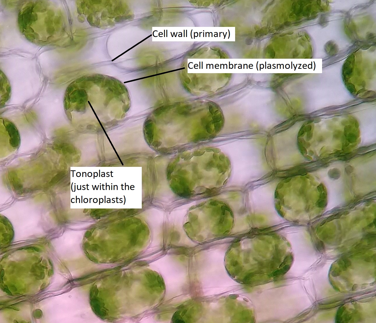
A bacteria diagram basically facilitates us to profit extra about this unmarried cell organisms that have … At hydrilla leaf when observed under a microscope, clearly visible cell wall, cell membrane, cytoplasm chloroplasts and flow, the flow in the rotation, the movement occurs regularly and lasts dialam cell, the cytoplasm flow can be seen in the movement of chloroplasts. Observe the specimen under the microscope. Place the coverslip on the leaf. Elodea are common freshwater aquarium plants. Elodea leaf cell illustration from a microscope slide a drop of 10. Ppt comparing animal and plant cell microscope lab powerpoint presentation id 842712. Figure 1 effects of hypertonic, isotonic and hypotonic solutions on plant cells. Single cell c4 photosynthesis in aquatic and terrestrial plants a. See how a generalized structure of an animal cell and plant cell look with labeled diagrams. Plant cell diagram under microscope. Cell is a tiny structure and functional unit of a living organism containing various parts known as organelles. Here's a diagram of a plant cell:
For plant cells, there is a cell wall. The diagram is very clear, and labeled; Play the video of elodea leaf cells with structures labeled so students can check their understanding. To prepare a sample for observation, slice a thin layer off an elodea leaf, place it on a glass slide and add a drop of water. Draw a few water plant cells below, labeling the nucleus, cytoplasm, cell wall, and the
3 from Elodea are common freshwater aquarium plants. Dicotyledon root.corte transversal de sarsaparrilla.140x. Lab manual exercise 1 lab manual exercise 1 virtual biology labs study botany lab practical flashcards. Observe the specimen under the microscope. The cell wall is very prominent under the microscope. 13 best fashion seasons cells images plant cell things under a. Place the leaf or algae sample on a slide and add. Always use one hand around the microscope arm and one hand under the microscope base.
Labelled diagram of a plant cell under a microscope.
Lec 2 plant cell structures. 40 new animal cell under electron. Actin based photo orientation movement of chloroplasts in plant. The microscope is used for looking at many specimens that cannot be seen with the… Do an epidermal peel (as per leaf epidermal peel practical, and perform this experiment. Elodea are common freshwater aquarium plants. First find water plant cells using 4x objective, then change to 10x and focus and draw, then turn to 40x and draw. Lab manual exercise 1 lab manual exercise 1 virtual biology labs study botany lab practical flashcards. Leaf structure under the microscope. Cell structure hydrilla, view of the leaf surface showing plant cells under the microscope. Ppt comparing animal and plant cell microscope lab powerpoint presentation id 842712. Place the microscope gently on the lab bench with the arm toward you. To do this a compound microscope is required given that it allows for higher magnification.
To prepare a sample for observation, slice a thin layer off an elodea leaf, place it on a glass slide and add a drop of water plant cell under microscope labeled. The diagram is very clear, and labeled;